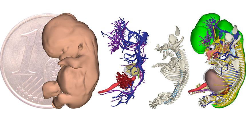
Database provides a rare peek at a human embryo’s first weeks

When I first found out my daughter existed, she was about half the size of a mini chocolate chip.
I was six weeks pregnant; she was four weeks into development. (The pregnancy timer officially begins two weeks before conception.) Already, the structures that would become her eyes had formed rudimentary orbs and the four tiny chambers of her heart were taking shape. At this stage of development, the embryo’s heart is huge, like a dumpling squeezed inside the torso.
You can see this early human heart and what happens before and after it develops with a new tool, the 3-D Atlas of Human Embryology, published November 25 in Science. The atlas chronicles the very first stages of human development — when growth is literally exponential and an embryo is building bodily systems that will be in place for a lifetime.
Embryologist Bernadette de Bakker and colleagues at the Academic Medical Center in Amsterdam created the atlas to help students, doctors and researchers better understand what goes on in those earliest weeks.
“We might know more about the moon than about our own development,” de Bakker says. Even today, human embryology textbooks often rely on pictures of chick or mouse embryos to describe how humans grow. Any human embryonic data used “is often based on just one or two specimens,” she says.
And that’s a problem because, as de Bakker has discovered, not all human embryos are the same.
Her team photographed nearly 15,000 cross sections of human embryos from the Carnegie Collection, a famous set of historical specimens collected from hysterectomies and abnormal pregnancies or miscarriages. De Bakker and colleagues uploaded the photos into a computer program, and then, using digital pencils and drawing pads, traced and labeled every organ and structure in every photo. It took some 75 people roughly 45,000 hours to complete.
What they (and we) have gained is a remarkable look at humans’ first metaphorical steps — the steady developmental march that, eventually, takes an embryo from a bundle of cells to babyhood.
Here are some landmarks in the first 60 days of development (links are to PDFs; save and open in Adobe Reader X or higher for interactive features):
Days 15-17: The embryo and all the membranes that surround it are no bigger than a speck of dust. The embryo itself is a speck within the speck, and consists of just three layers of cells. From these layers, all organs and body structures will form.
Days 19-21: What a difference a few days make. The embryo is now about the size of a pinhead and has laid the early groundwork for the heart, gut, skin, muscles, skeleton and brain.
Days 21-23: A furrow of tissue that gives rise to the brain and spinal cord has now begun to fold together, forming the neural tube. If the furrow doesn’t close properly, the spinal cord could protrude from the backbone, a birth defect called spina bifida.
Days 28-32: The embryo, now at the half mini-chocolate-chip size, starts to take on a textbook look, fledgling head and lower body curled toward each other — a rough draft of the classic fetal position.
Days 35-38: Short paddlelike arms and the first nubs of legs have emerged. The embryo is now almost the size of a ladybug.
Days 44-48: It’s now a slightly bigger ladybug, and leg bones and hand and finger bones have formed. The liver, suddenly, has ballooned in size, filling much of the lower half of the body cavity.
Days 51-53: Chubby fingers have sprouted from once clublike hands. Toes are not visible yet, but toe bones are in place. The embryo is roughly marble-sized.
Days 56-60: The embryonic brain is a giant bulbous globe now, more than a third of the entire body — which is about the size of a cherry tomato. Skinny arms and legs fold close, and a plate of skull has started to stretch across the back of the head.
Give it another 30 weeks or so and that tiny embryo will grow to the size of a bowling ball. Comparatively, a little chocolate chip doesn’t seem like much. But development-wise, it goes through something pretty huge.