A message to rock climbers: Be kind to nature
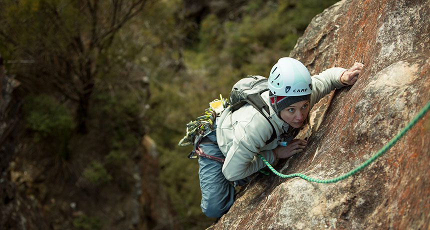
For the millions of people who have taken up the sport of rock climbing, a cliff face is a challenge, a vertical puzzle solved only with the proper placement of hands and feet. Look closely, though, and those crevices and cracks that provide hand- and footholds also provide homes for a variety of plants, invertebrates and other easily overlooked species.
People who participate in outdoor sports like rock climbing may not think about the environmental impact of what they’re doing. After all, how big of an impact can one person really have on a rock? But there is a potential for harm, notes ecologist Andrea Holzschuh of the University of Würzburg in Germany. Finding evidence of that harm, though, is a challenge — the features that make some cliffs fun to climb, or not, also make for complicated research.
Holzschuh became interested in the effects that climbers are having on the environment in part because she is a climber herself, partial to tackling rocks in the Frankenjura region of Germany, which is noted for having some of the best climbing in Europe. The plants, animals and other species that make the cliffs their home, she notes, are often specialists that have found ways to adapt and even thrive in the extreme conditions found on the rock face. They may be rare or completely absent from nearby spots, and they often are slow-growing and their numbers grow only in spurts.
And then come the climbers, who may trample what grows at the bottom of a cliff, dig out whatever is growing in a crevice to gain a better handhold, spread species not native to the area or taint the rock face with chalk, altering pH or nutrient conditions for whatever is growing there. Rock climbing isn’t quite as impact-free as some might assume.
But scientists haven’t really been able to adequately assess that impact. Holzschuh went looking for research on this topic and found only 22 studies that tested how rock climbing might affect plants or animals. She tossed out six of those studies because they failed to make comparisons with unclimbed areas or had other major design problems that made it impossible to tease out effects. The remaining 16 studies found a variety of impacts on organisms ranging from lichens to snails to cedar trees. Holzschuh’s review appears in the December Biological Conservation.
But what the review really highlights is just how difficult it is to study rock climbing’s potential impacts. Holzschuh says a big challenge is in finding appropriate unclimbed cliff faces to compare to those that rock climbers frequent — ones that share traits such as slope and how much sunlight the face gets. “Often, all cliffs in a regions that are attractive for climbers are climbed and only cliffs that do not resemble the climbed cliffs in all abiotic traits remain unclimbed,” she notes. “Then no reliable study can be conducted.”
And then, of course, there’s the inaccessibility of many cliffs and the difficulty in studying even the accessible ones. “How many people have these skills and the flexibility to work on these projects?” says Michael Tessler of the American Museum of Natural History and Fordham University. Plus, he notes that a subset of rock climbing called bouldering — in which climbers tackle boulders or short cliff faces measuring less than 3.5 meters high, without using safety ropes — is especially popular with younger people. “Professors inherently aren’t always young,” he notes.
Tessler and colleague Theresa Clark of the University of Nevada, Las Vegas published the first ever analysis attempting to quantify the impact of bouldering on the environment. This type of climbing has similar potential for ecosystem damage as roped rock climbing, they note, plus a couple of additional ones: Boulderers often clear the ground below of rocks and logs so that they can place crash pads in case of falls, and they may be more likely to trample anything at the top of a boulder or cliff, rather than coming directly down.
Tessler and Clark tried to measure the impact of climbers at bouldering routes in the Shawangunk Ridge, a popular climbing site in New York where Tessler climbs. They compared transects in climbed boulder routes with transects along nearby unclimbed sections of rock and found differences in lichen, moss and woody plants. None of this added up to a major threat, but conservation managers might want to monitor these activities in remote sites and shut down certain routes that are proving too popular — and potentially too harmful to whatever is growing there, Tessler and Clark suggest in the December Biological Conservation.
While we still can’t really say how much impact climbers might be having on the rocky environments they climb, there is a definite need for more scientists to strap on their climbing shoes and tackle the questions of climbing’s impact. (Try it! It’s lots of fun!) But climbers, too, can do their part, Holzschuh and Tessler say.
“I think climbers can easily minimize their impact on the cliff vegetation if they do not willingly remove vegetation from the cliff to ‘clean’ hand- and footholds in the climbing route. Climbers should not access the cliff plateau [and should] leave this cliff part completely undisturbed,” Holzschuh says. “At the cliff base, bags and gear should be laid down within a small area to reduce the effects of trampling.”
Tessler also has advice. “Boulderers should be aware that even infrequent climbing leaves some impression on rock-associated vegetation,” he says. “They should remove as little vegetation and soil when climbing and establishing climbs. Also, if a climb is wet, dirty or covered in vegetation, maybe go to another one. This is an easy way to ensure that some rock faces can stay more natural.”
And if climbing is restricted because, say, rare birds are breeding there, rock climbers should obey the restriction and go climb somewhere else, Holzschuh says. There are plenty of other cliffs to be conquered.