Genes could record forensic clues to time of death
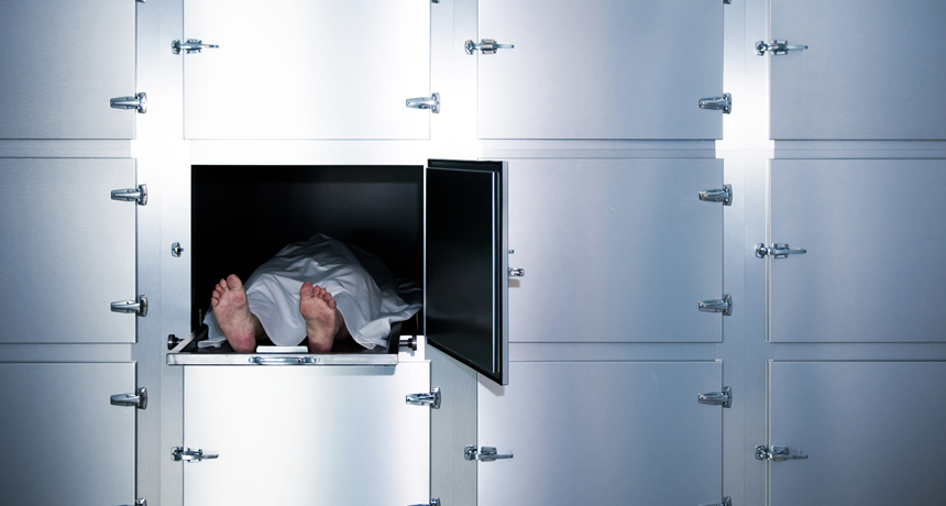
Dying, it turns out, is not like flipping a switch. Genes keep working for a while after a person dies, and scientists have used that activity in the lab to pinpoint time of death to within about nine minutes.

During the first 24 hours after death, genetic changes kick in across various human tissues, creating patterns of activity that can be used to roughly predict when someone died, researchers report February 13 in Nature Communications.
“This is really cool, just from a biological discovery standpoint,” says microbial ecologist Jennifer DeBruyn of the University of Tennessee in Knoxville who was not part of the study. “What do our cells do after we die, and what actually is death?”

What has become clear is that death isn’t the immediate end for genes. Some mouse and zebrafish genes remain active for up to four days after the animals die, scientists reported in 2017 in Open Biology.
In the new work, researchers examined changes in DNA’s chemical cousin, RNA. “There’s been a dogma that RNA is a weak, unstable molecule,” says Tom Gilbert, a geneticist at the Natural History Museum of Denmark in Copenhagen who has studied postmortem genetics. “So people always assumed that DNA might survive after death, but RNA would be gone.”
But recent research has found that RNA can be surprisingly stable, and some genes in our DNA even continue to be transcribed, or written, into RNA after we die, Gilbert says. “It’s not like you need a brain for gene expression,” he says. Molecular processes can continue until the necessary enzymes and chemical components run out.

“It’s no different than if you’re cooking a pasta and it’s boiling — if you turn the cooker off, it’s still going to bubble away, just at a slower and slower rate,” he says.

No one knows exactly how long a human’s molecular pot might keep bubbling, but geneticist and study leader Roderic Guigó of the Centre for Genomic Regulation in Barcelona says his team’s work may help toward figuring that out. “I think it’s an interesting question,” he says. “When does everything stop?”

Tissues from the dead are frequently used in genetic research, and Guigó and his colleagues had initially set out to learn how genetic activity, or gene expression, compares in dead and living tissues.

The researchers analyzed gene activity and degradation in 36 different kinds of human tissue, such as the brain, skin and lungs. Tissue samples were collected from more than 500 donors who had been dead for up to 29 hours. Postmortem gene activity varied in each tissue, the scientists found, and they used a computer to search for patterns in this activity. Just four tissues, taken together, could give a reliable time of death: subcutaneous fat, lung, thyroid and skin exposed to the sun.

Based on those results, the team developed an algorithm that a medical examiner might one day use to determine time of death. Using tissues in the lab, the algorithm could estimate the time of death to within about nine minutes, performing best during the first few hours after death, DeBruyn says.

For medical examiners, real-world conditions might not allow for such accuracy.

Traditionally, medical examiners use body temperature and physical signs such as rigor mortis to determine time of death. But scientists including DeBruyn are also starting to look at timing death using changes in the microbial community during decomposition (SN Online: 7/22/15).

These approaches — tracking microbial communities and gene activity — are “definitely complementary,” DeBruyn says. In the first 24 hours after death, bacteria, unlike genes, haven’t changed much, so a person’s genetic activity may be more useful for zeroing in on how long ago he or she died during that time frame. At longer time scales, microbes may work better.

“The biggest challenge is nailing down variability,” DeBruyn says. Everything from the temperature where a body is found to the deceased’s age could potentially affect how many and which genes are active after death. So scientists will have to do more experiments to account for these factors before the new method can be widely used.
Cutting off a brain enzyme reversed Alzheimer’s plaques in mice

Knocking back an enzyme swept mouse brains clean of protein globs that are a sign of Alzheimer’s disease. Reducing the enzyme is known to keep these nerve-damaging plaques from forming. But the disappearance of existing plaques was unexpected, researchers report online February 14 in the Journal of Experimental Medicine.

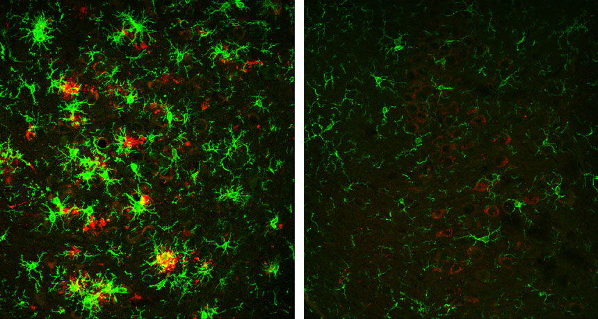
The brains of mice engineered to develop Alzheimer’s disease were riddled with these plaques, clumps of amyloid-beta protein fragments, by the time the animals were 10 months old. But the brains of 10-month-old Alzheimer’s mice that had a severely reduced amount of an enzyme called BACE1 were essentially clear of new and old plaques.
Studies rarely demonstrate the removal of existing plaques, says neuroscientist John Cirrito of Washington University in St. Louis who was not involved in the study. “It suggests there is something special about BACE1,” he says, but exactly what that might be remains unclear.

Story continues below graphic
One theory to how Alzheimer’s develops is called the amyloid cascade hypothesis. Accumulation of globs of A-beta protein bits, the idea goes, drives the nerve cell loss and dementia seen in the disease, which an estimated 5.5 million Americans had in 2017. If the theory is right, then targeting the BACE1 enzyme, which cuts up another protein to make A-beta, may help patients.
BACE1 was discovered about 20 years ago. Initial studies turned off the gene that makes BACE1 in mice for their entire lives, and those animals produced almost no A-beta. In humans, however, any drug that combats Alzheimer’s by going after the enzyme would be given to adults. So Riqiang Yan, one of the discoverers of BACE1 and a neuroscientist at the Cleveland Clinic, and colleagues set out to learn what happens when mice who start life with normal amounts of BACE1 lose much of the enzyme later on.

The researchers studied mice engineered to develop plaques in their brains when the animals are about 10 weeks old. Some of these mice were also engineered so that levels of the BACE1 enzyme, which is mostly found in the brain, gradually tapered off over time. When these mice were 4 months old, the animals had lost about 80 percent of the enzyme.
Alzheimer’s mice with normal BACE1 levels experienced a steady increase in plaques, clearly seen in samples of their brains. In Alzheimer’s mice without BACE1, however, the clumps followed a different trajectory. The number of plaques initially grew, but by the time the mice were around 6 months old, those plaques had mostly disappeared. And by 10 months, “we hardly see any,” Yan says.

Cirrito was surprised that getting rid of BACE1 later in life didn’t just stop plaques from forming, but removed them, too. “It is possible that perhaps a therapeutic agent targeting BACE1 in humans might have a similar effect,” he says.

Drugs that target BACE1 are already in development. But the enzyme has other jobs in the brain, such as potentially affecting the ability of nerve cells to communicate properly. It may be necessary for a drug to inhibit some, but not all, of the enzyme, enough to prevent plaque formation but also preserve normal signaling between nerve cells, Yan says.

A new study eases fears of a link between autism and prenatal ultrasounds

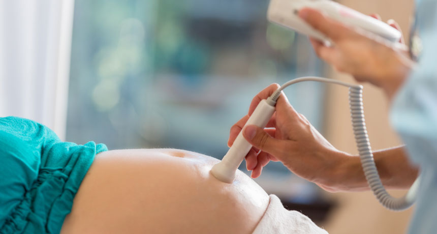
Ultrasounds during pregnancy can be lots of fun, offering peeks at the baby-to-be. But ultrasounds aren’t just a way to get Facebook fodder. They are medical procedures that involve sound waves, technology that could, in theory, affect a growing fetus.

With that concern in mind, some researchers have wondered if the rising rates of autism diagnoses could have anything to do with the increasing number of ultrasound scans that women receive during pregnancy.

The answer is no, suggests a study published online February 12 in JAMA Pediatrics. On average, children with autism were exposed to fewer ultrasounds during pregnancy, scientists found. The results should be “very reassuring” to parents, says study coauthor Jodi Abbott, a maternal fetal medicine specialist at Boston Medical Center and Boston University School of Medicine.
To back up: Autism rates have risen sharply over the last several decades (though are possibly plateauing). Against this backdrop, researchers are searching for the causes of autism — and there are probably many. Autism is known to run in families, and scientists have found some of the particular genetic hot spots that may contribute. Other factors, such as older parents and maternal obesity, can also increase the risk of autism.

Scientists suspect that in many cases, autism is caused by many factors, all working together. Could prenatal ultrasounds, which have become more routine and more powerful, be one of those factors? These scans use sound waves that penetrate mothers’ bodies, and then collect the waves that bounce back, forming a picture of fetal tissues. During this process, the waves may be able to heat up the tissue they travel through.

Work on animals has suggested that ultrasounds can in fact interfere with fetal brain development, derailing the normal movements of cells that populate the brain. Mice exposed to 30 or more minutes of ultrasound in utero had abnormal brain development, for instance. But it’s not at all clear whether a similar thing might happen in humans, and if so, whether such effects might contribute to autism.
The new study compared ultrasound exposure among three groups: 107 children diagnosed with autism spectrum disorder, 104 children diagnosed with a developmental delay, and 209 typically developing children. On average, the children with autism were exposed to 5.9 ultrasound scans over the course of pregnancy. Children with developmental delays were exposed to 6.1 scans, and typically developing children were exposed to 6.3 scans, the researchers found. (For all groups, these numbers are way above the one to two scans per low-risk pregnancy recommended by the American College of Obstetricians and Gynecologists.)

For all three groups, the duration of the scans was similar. So was the thermal index, an indication of how much warming might have happened. “In almost every parameter we looked at, ultrasound seemed perfectly safe,” says study coauthor N. Paul Rosman, a pediatric neurologist at Boston Medical Center and Boston University School of Medicine.

One measure was different, the researchers found: During the first trimester, mothers who had children with autism had slightly deeper ultrasounds than women who had typically developing children and children with developmental delays. Ultrasound depth measures the distance from the transducer paddle that emits the waves to the spot that’s being imaged. The measure “has a lot to do with the size of the mother and the distance between her skin, where the ultrasound transducer is, and where the baby is,” Abbott says.

Lots of questions remain about whether — and how — ultrasound depth, or other aspects of the technology, might affect fetuses. “The study certainly wasn’t perfect,” Rosman says. It combed back through medical records of women instead of following women from the beginning. And it didn’t control for certain traits that may influence autism, such as smoking.

The results suggest that on their own, ultrasounds don’t cause autism spectrum disorder, says Sara Jane Webb of Seattle Children’s Research Institute and the University of Washington, who cowrote a JAMA Pediatrics companion piece. “At this time, there is no evidence that ultrasound is a primary contributor to poor developmental outcomes when delivered within medical guidelines,” she says.

While there’s more science to sort out here, the news is reassuring for women who might be worried about getting scanned. Women should follow their doctors’ guidance on ultrasounds, Rosman says. “We don’t think there’s anything in this study to recommend otherwise.”

A new species of tardigrade lays eggs covered with doodads and streamers

What a spectacular Easter basket tardigrade eggs would make — at least for those celebrating in miniature.

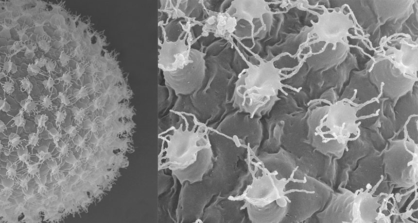
A new species of the pudgy, eight-legged, water creatures lays pale, spherical microscopic eggs studded with domes crowned in long, trailing streamers.

Eggs of many land-based tardigrades have bumps, spines, filaments and such, presumably to help attach to a surface, says species codiscoverer Kazuharu Arakawa. The combination of a relatively plain surface on the egg itself (no pores, for instance) plus a filament crown helps distinguish this water bear as a new species, now named Macrobiotus shonaicus, he and colleagues report February 28 in PLOS ONE.
With about 20 new species added each year to the existing 1,200 or so known worldwide, tardigrades have become tiny icons of extreme survival (SN Online: 7/14/17).

“I was actually not looking for a new species,” Arakawa says. He happened on it when searching through moss he plucked from the concrete parking lot at his apartment. He routinely samples such stray spots to search for tardigrades, one of his main interests as a genome biologist at Keio University’s Institute for Advanced Biosciences in Tsuruoka City, Japan.
These particular moss-loving creatures managed to grow and reproduce in the lab —“very rare for a tardigrade,” he says. He didn’t realize it was an unknown species until he started deciphering the DNA that makes up some of its genes. The sequences he found didn’t match any in a worldwide database.

His two coauthors, at Jagiellonian University in Krakow, Poland, worked out that he had found a new member of a storied cluster of relatives of the tardigrade M. hufelandi. That species, described in 1834, kept turning up across continents around the world — or so biologists thought for more than a century. Realization eventually dawned that the single species that could live in such varied places was actually a complex of close cousins.

And now M. shonaicus adds yet another cousin to a group of about 30. Who knows where the next one will turn up. “I think there are lots more to be identified,” Arakawa says.

The debate over how long our brains keep making new nerve cells heats up

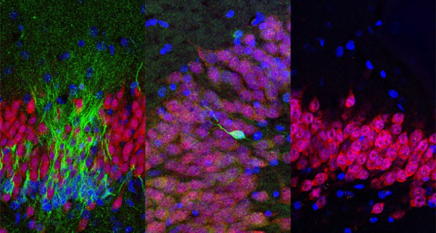
Adult mice and other rodents sprout new nerve cells in memory-related parts of their brains. People, not so much. That’s the surprising conclusion of a series of experiments on human brains of various ages first described at a meeting in November (SN: 12/9/17, p. 10). A more complete description of the finding, published online March 7 in Nature, gives heft to the controversial result, as well as ammo to researchers looking for reasons to be skeptical of the findings.

In contrast to earlier prominent studies, Shawn Sorrells of the University of California, San Francisco and his colleagues failed to find newborn nerve cells in the memory-related hippocampi of adult brains. The team looked for these cells in nonliving brain samples in two ways: molecular markers that tag dividing cells and young nerve cells, and telltale shapes of newborn cells. Using these metrics, the researchers saw signs of newborn nerve cells in fetal brains and brains from the first year of life, but they became rarer in older children. And the brains of adults had none.

There is no surefire way to spot new nerve cells, particularly in live brains; each way comes with caveats. “These findings are certain to stir up controversy,” neuroscientist Jason Snyder of the University of British Columbia writes in an accompanying commentary in the same issue of Nature.