In a study of pregnant women in Brazil, nearly 30 percent of those infected with Zika virus had babies with fetal abnormalities, researchers report March 4 in the New England Journal of Medicine.
Zika virus is the leading suspect for what’s causing a spike in certain birth defects reported in Brazil. Scientists have previously found traces of Zika in the brains of fetuses with microcephaly (a birth defect that leaves babies with smaller-than-normal heads). And one study has reported that the virus can infect and kill a cell type crucial to developing brains (SN Online: 3/4/16).
The new study enrolled 88 pregnant women from Rio de Janeiro who had developed a rash (a sign of Zika infection). They tracked the women throughout their pregnancies; so far, eight have given birth. Of the 42 women who both tested positive for Zika and received fetal ultrasounds, 12 of the women’s babies had abnormalities (including small heads, damaged brain tissue, and low levels of amniotic fluid).
Despite mild clinical symptoms, Zika infection during pregnancy appears to be linked with grave outcomes, the authors write.
Asteroids probably ferried water to an infant Earth (SN: 5/16/15, p. 18), but they weren’t responsible for our planet’s entire chemical inventory. Comets might have trucked in noble gases and much of the raw material needed for life, researchers suggest online March 9 in Earth and Planetary Science Letters.
Noble gases don’t play well with the other elements. They typically shun chemical reactions, which means their abundances haven’t changed much since the formation of the solar system. Comets are thought to be frozen relics from the birth of the planets, but until recently researchers didn’t know how much of any noble gas comets carried. That changed shortly after the Rosetta spacecraft arrived at comet 67P/Churyumov-Gerasimenko in August 2014 (SN: 9/6/14, p. 8) and made the first measurement of argon in a cometary atmosphere. It’s not much — roughly 1/100,000 the amount of water — but it’s several orders of magnitude more than the abundance of argon in icy asteroids. And that’s enough for comets to have been a major source of argon (and presumably other noble gases) to Earth, Bernard Marty, a geochemist at Petrographic and Geochemical Research Centerin Vandœuvre-lès-Nancy, France, and colleagues report.
The delivery arrived during the Late Heavy Bombardment about 600 million years after the start of the solar system, Marty and collaborators suggest. That’s when the moon (and supposedly Earth) was pummeled by debris from the outer solar system as the giant planets abruptly settled into their current orbits.
Argon might seem of little relevance to everyday life. But “if argon came from comets, we can make some inferences about how much prebiotic material came in comets too,” Marty says. And those other goodies, such as amino acids, might matter a great deal. Assuming that all the argon in Earth’s atmosphere came from comets, and that cometary levels of amino acids (which no one has measured) are similar to what’s found in meteorites known as carbonaceous chondrites, the researchers calculated how much amino acids comets could have delivered to Earth. Dust collected from comet 81P/Wild 2 by the Stardust spacecraft in 2004 showed some chemical similarities between its quarry and those meteorites.
It’s a rough calculation, Marty admits, and it assumes that the deliveries survived the impacts. But the team estimates that the amount of amino acids supplied by comets could roughly equal the total mass found in all organisms from paramecia to plants and people.
“I think it’s an interesting exercise,” says Conel Alexander, a planetary scientist at the Carnegie Institution for Science in Washington, D.C. “But it’s full of so many uncertainties. My worries are that we still know so little about comet composition.” The amount of cometary argon, for example, is based on just one comet. Other comets have shown tremendous variability, for example, in the relative amounts of water isotopes. Researchers also don’t have a good handle on the concentration of other noble gases, such as xenon, lurking in comets. There’s also uncertainty about the Late Heavy Bombardment that presumably brought the comets to Earth. Evidence for the influx of debris comes from a spike in craters on the moon roughly 4 billion years ago. But the timing comes from lunar rocks collected by Apollo astronauts and those samples might all have come from one basin, says Alexander. Rather than revealing the ages of many craters, the moon rocks might record the date of a single run-in with a giant intruder.
Recent computer simulations also indicate that the giant planet tango that allegedly triggered the bombardment should have removed at least one of the inner planets, which doesn’t appear to have happened. To avoid that catastrophe, the gas giants had to have settled down before the rocky planets finished forming and so wouldn’t have been available to fling things at Earth 600 million years later.
Bacteria is back, baby. After decades of gobbling antibiotics and overzealous hand sanitizing, it’s now clear that the bacteria that live in and on our bodies can help keep us healthy. That realization is what led scientists to rub brand-spanking-new babies with fluid from the mothers’ vaginas.
Like about a third of babies born in the United States, these babies were born by C-section, and so missed out on a trip through the birth canal, where their bodies would have been propelled with viselike pressure through a channel coated with microbe-laden fluid. This crushing, juicy journey coats babies with their mothers’ vaginal microbes. Babies born by C-section are instead colonized with bacteria that live on skin (possibly picked up from the dust in the hospital operating room).
That difference may have important implications for future health, some scientists think. Studies have hinted that microbes picked up during a vaginal birth can sculpt newborns’ immune systems in ways that combat disorders including obesity, asthma and allergies. So it follows that replacing those missing vaginal microbes might be a good thing.
Scientists led by Maria Dominguez-Bello of New York University and the University of Puerto Rico in San Juan took a first step in testing that idea with the vaginal wipe-down experiment. An hour before a C-section, doctors inserted a square of wet, folded gauze into four women’s vaginas to slurp up the fluid. This microbe-laden gauze came out right before the C-sections began. Within a minute of birth, the newborns were swabbed with the gauze, first on their lips, then their faces, trunks, arms and legs, genitals, anuses and, finally, their backs. The whole-body rubdown took about 15 seconds.
Those 15 seconds led to bacterial changes that lasted through the newborns’ first month, the researchers reported February 1 in Nature Medicine. Compared with babies born via C-section who didn’t get swabbed, the four swabbed babies had bacterial species on their mouths, skin and guts that were more similar to those in their mothers’ vaginas. That resemblance suggests that swabbing could transform the newborns’ microbes in a way that might ultimately be beneficial.
The study gives some much-needed heft to the idea that microbes matter. By showing that newborns’ bacteria can be manipulated in a pretty simple way, the study opens the door for other tests of whether this microbial rehab is a good thing.
But the study is preliminary, the authors stress in their paper. The results come from four babies, with only a month of follow-up. It’s possible that these changes don’t stick around. It’s also possible that these microbes don’t actually improve health.
Those outstanding questions haven’t deterred some intrepid parents of babies born by C-section who want to try “vaginal seeding,” says pediatric infectious disease expert Aubrey Cunnington of Imperial College London. Over the last several years, news reports have raised interest, prompting some parents to request the procedure. On neonatal infection rounds last summer, a fellow doctor brought up a troubling story. “She described a recent situation where she had needed to stop a midwife from performing seeding, because the mother had a genital herpes infection,” Cunnington says.
That situation raised an important issue — fluid from a mother’s vagina may carry beneficial microbes, but it could also hold bacteria and viruses that could harm a newborn, Cunnington and colleagues wrote in an editorial published February 23 in BMJ. “Demand has outstripped both professional awareness and professional guidance on this practice,” he and his colleagues wrote in their editorial.
A lack of guidance is worrisome, he argues, because the procedure could unknowingly expose newborns to dangerous bugs, pathogens that babies born by C-section usually avoid. Group B streptococcus, which is carried by about 30 percent of women, can trigger meningitis and fatal septicemia, he says. Herpes simplex virus can lead to death and disability in newborns. And chlamydia and gonorrhea can cause severe eye infections.
Cunnington argues that those potential risks, coupled with unproven and potentially slim benefits, makes the procedure a no-go for now. “On balance, I don’t think the potential benefit outweighs the risk,” he says. Health practitioners at his hospital have been advised not to perform the procedure. (But because the swabbing is so simple, they can’t stop parents, or more realistically, another helper, from performing it themselves.) Staff at other hospitals vary in their willingness to help.
In the swabbing study, the protocol came with built-in safeguards. The women were tested for pathogens, and showed no signs of viral or bacterial infections. And the gauze was handled carefully so that it didn’t pick up new germs. Those precautions should be followed by any hospital or DIYer.
Like any decision — particularly those related to pregnancy and children — choosing whether to swab a baby born by C-section comes with a murky risk assessment. Some of that risk can be lowered by ruling out pathogens such as group B strep and STDs, tests that are usually offered to pregnant women in the United States. Cunnington points out that group B strep test results aren’t always reliable, and that STDs can be picked up after the tests. As a result, looking for those pathogens close to the time of delivery can make microbe swabbing less risky.
If you don’t feel like an adventurous self-experimenter, you can tend to your newborn’s microbes in other ways. Breastfeeding and avoiding unnecessary antibiotics may both encourage good microbes to flourish, no swabbing necessary.
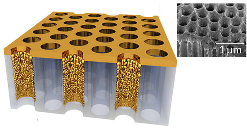
A new, extremely black material can turn water into steam using only sunlight, without the need to bring the water to a boil. Made of gold nanoparticles tens of billionths of a meter wide affixed to a scaffold pocked with tiny channels, or “nanopores,” the material is a deep black color because it reflects very little visible light. It is 99 percent efficient at absorbing light in the visible spectrum and parts of the infrared spectrum, researchers report April 8 in Science Advances.
Thanks to its highly porous structure, the material floats on the surface of water, allowing it to soak up the sun’s rays. When light of a certain wavelength hits a gold nanoparticle inside one of the nanopores, it stirs up the electrons on the surface, sloshing them back in forth in an oscillation known as a plasmon. These plasmons produce localized, intense heating, which vaporizes the water nearby. The wavelength of light that excites a plasmon depends on the size of the nanoparticle. So in order to take advantage of as much of the sun’s output as possible, the group interspersed a variety of sizes of gold nanoparticles in the pores, which could therefore absorb a range of wavelengths. It’s not the first time scientists have produced steam with plasmonic materials, but the new material improves the efficiency of the process, converting up to 90 percent of the light’s energy into steam, says materials scientist Jia Zhu of Nanjing University in China, a leader of the research group.
“They have really come out with a very intriguing solution,” says mechanical engineer Nicholas Fang of MIT, who was not involved in the research. The efficiency isn’t quite as high as scientists have achieved with certain other types of materials, like carbon nanotubes, Fang says. But the new material should be cheaper to manufacture.
Efficient steam generation could be useful for desalination, producing freshwater from salty water, says Zhu. Other potential applications range from sterilization to running steam engines. “Steam can be used for many other things,” he says. “It is a very useful form of energy.”
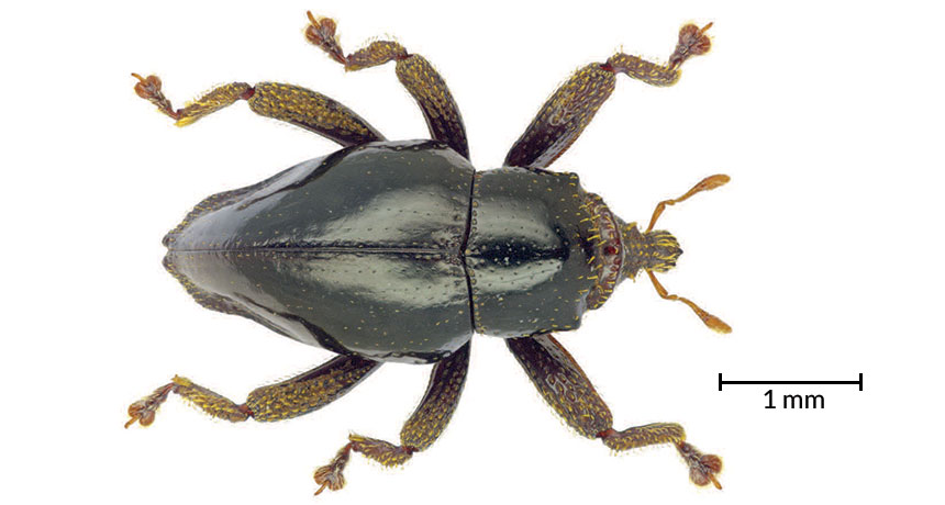
In a galaxy far, far away, Chewbacca is a 7.5-foot-tall Wookiee. On Earth, he’s a small furry beetle.
Researchers discovered four new species of weevils on an island off the coast of Papua New Guinea, one of which they named after the lofty Star Wars character. Trigonopterus chewbacca is a black, flightless beetle about 3 millimeters long that thrives in the tropical forests of New Britain. Although T. chewbacca doesn’t resemble its namesake in size, the dense hairlike scales covering its head and legs reminded the researchers of Chewbacca’s fur.
Before these finds, Trigonopterus beetles hadn’t been spotted on New Britain. The discovery of T. chewbacca and its three relatives, T. obsidianus, T. puncticollis and T. silaliensis, suggests that the genus colonized the island at least four separate times, the team reports April 21 in ZooKeys.
T. chewbacca joins the ranks of other insects with a Star Wars moniker. Among its peers: a furry moth also named after the heroic Wookiee, a wasp named for Yoda and a Darth Vader slime-mold beetle.
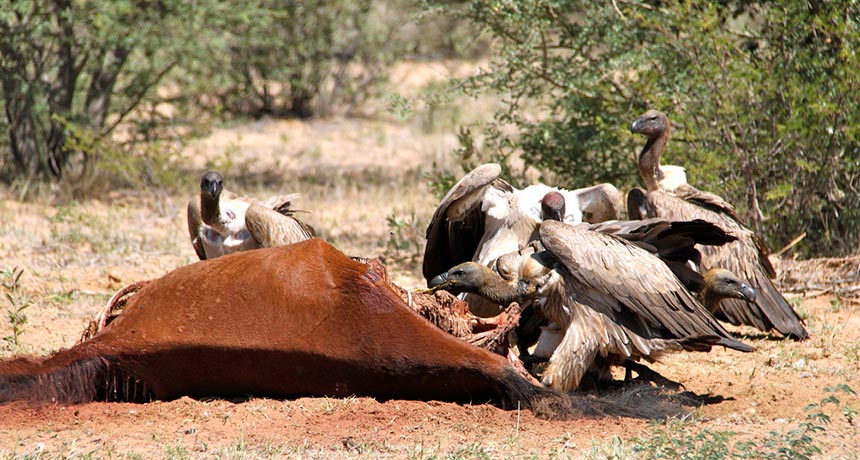
Vultures are the birds everyone loves to hate. Even though you have nothing to fear from them — unless you’re dead — vultures’ steady diet of carrion will gross most people out. That diet may also be responsible for the birds’ quick and steep declines around the globe, a new study shows.
It’s not the dead bodies that are killing vultures, though. It’s the poisons with which humans have laced those meals, both intentionally and inadvertently, Evan Buechley and Çağan Şekercioğlu of the University of Utah in Salt Lake City conclude in the June Biological Conservation.
The team went searching for an explanation to something Şekercioğlu had reported in 2004 and is still true today — that vultures are the most threatened group of birds. Of the 22 species of vultures, nine are now critically endangered, three are endangered and four are near threatened, according to the International Union for Conservation of Nature, which tallies endangered species.
Buechley and Şekercioğlu were looking for an explanation of why these scavenging species (called “obligate scavengers” because they depend almost entirely on carrion for survival) are doing so poorly but “facultative scavengers” — birds such as storks, gulls and crows that can also eat things other than carrion and trash — tend to be doing well and even increasing in numbers in many cases. The researchers collected ecological information and population trend data on the 22 species of vultures and other avian scavengers and then tried to figure out what made the vultures so vulnerable.
Some aspects of biology do contribute to the vulture declines, the team found. These are large animals that live long and don’t produce a lot of offspring. That means that populations can take a long time to recover from bird deaths. But the ultimate cause of those deaths is what is disturbing — dietary toxins, which are the primary cause of declines in 14 of the 16 threatened and near-threatened vulture species, the team found.
Those toxins come in various forms. In India and Southeast Asia, it’s the cattle drug diclofenac, which causes kidney failure in any vulture unlucky enough to come across a cow that didn’t survive its medical treatment. Diclofenac is a problem for vultures in Africa, too, (and now Spain), but there the birds have also fallen victim to the poisons used to kill hyenas, jackals and lions in response to dead livestock. Wildlife poachers have also deliberately poisoned their prey in an effort to get rid of the circling vultures that can alert authorities to their crime. (Buechley and Şekercioğlu discovered a 2007 incident in Namibia in which a poisoned elephant carcass killed as many as 600 birds.) And in Europe and the Americas, carcasses laced with rodenticides, insecticides and lead from ammunition are also killing vulture species.
Without vultures, some of these ecosystems are already having problems. Other scavenging species aren’t quite able to fit into the vulture niche. They can’t eat as much and they don’t have stomachs equipped to kill deadly microbes, like vultures do. That means anything that does eat carrion could potentially spread disease. Populations of scavenging pests, like rats and feral dogs, have already skyrocketed in some places as these animals feast on what vultures would have once dealt with. Perhaps not surprisingly, that has led to problems, such as an increase in dog bites in India that has resulted in thousands of human deaths from rabies.
Much of the vulture declines could be easily solved by banning the chemicals that kill them, the researchers note. Because while vultures may be more inherently vulnerable to extinction than other bird species, due to their biology, their importance to the global ecosystem — and our own health — makes them too valuable to let slip away.
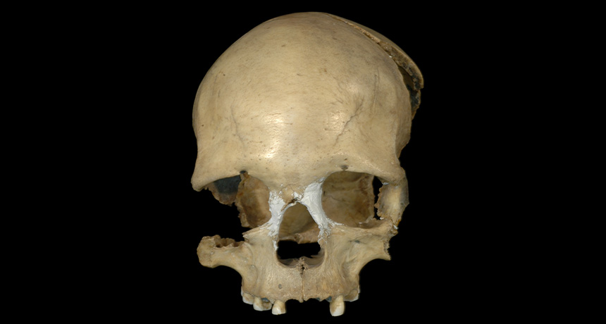
DNA from an ancient woman who lived in what’s now Romania indicates that people in Asia trekked to Africa starting between 45,000 and 40,000 years ago.
Evidence for this back-to-Africa trip comes from the partial remains of a 35,000-year-old Homo sapiens discovered in a Romanian cave more than 60 years ago. A distinctive pattern of alterations to mitochondrial DNA extracted from two of the teeth are similar to alterations seen in mitochondrial DNA of present-day North Africans, signaling an evolutionary connection, the team proposes May 19 in Scientific Reports.
After evolving in Africa around 200,000 years ago, human populations spread out of the continent by 50,000 years ago. The ancient Romanian woman’s DNA came from a maternal line that originated in West Asia after humans initially left Africa but then ended up in North Africa, the scientists propose.
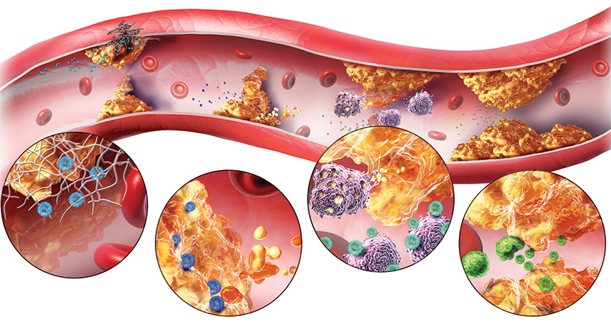
Careening through the bloodstream, a single nanoparticle is dwarfed by red blood cells whizzing by that are 100 times larger. But when specially designed nanoparticles bump into an atherosclerotic plaque — a fatty clog narrowing a blood vessel — the tiny particles can play an outsized role. They can cling to the plaque and begin to break it down, clearing the path for those big blood cells to flow more easily and calming the angry inflammation in the vicinity.
By finding and busting apart plaques in the arteries, nanoparticles may offer a new, non-surgical way to reduce a patient’s risk for heart attack and stroke.
Nanoparticles measure less than 100 nanometers across — a thousandth the thickness of a dollar bill. Despite being tiny, they can be engineered to haul a mix of molecules — such as tags that make them stick to a plaque, drugs that block inflammation or dyes that let scientists track their movements. Over the last two decades, scientists have exploited these strategies to fight cancer, designing nanoparticles that deliver drugs (SN Online: 1/3/14) or dyes for imaging deep into the core of a tumor. The U.S. Food and Drug Administration has approved a few dozen cancer-focused nanomedicines. Now researchers have begun engineering nanoparticles to target cardiovascular disease, which kills even more people each year than cancer. Nanosized compounds have been built that can sweep into clogged arteries to shrink the plaques that threaten to block blood flow. Some nanoparticles home in on the plaques by binding to immune cells in the area, some do so by mimicking natural cholesterol molecules and others search for collagen exposed in damaged vessel walls. Once at the location of a plaque, either the nanoparticles themselves or a piggybacked drug can do the cleanup work.
The aim of all these approaches is to prevent strokes and heart attacks in people with cardiovascular disease, either before surgery becomes necessary or after surgery to prevent a second event. Today, cardiovascular nanoparticles are still far from pharmacy shelves. Most have not reached safety testing in patients. But in mice, rats and pigs, nanodrugs have slowed the growth of the plaques that build up on vessel walls, and in some cases have been able to shrink or clear them.
“I think the effect we can have with these nanoparticles on cardiovascular disease is even more pronounced and direct than what we’ve seen in cancer,” says Prabhas Moghe, a biomedical engineer at Rutgers University in Piscataway, N.J. Every minute, more than a gallon of blood pumps through the human heart, pushing through miles of blood vessels to deliver oxygen and nutrients to organs and extremities. In a healthy person, the trip is as smooth as a drive on a freshly paved highway. But in the more than 10 percent of U.S. adults who have cardiovascular disease, the route might be more like a pothole-filled road squeezed by Jersey barriers.
Waxy globs, or plaques, of fat and cholesterol line the blood vessels, thickening and hardening the walls, impeding blood flow. As fat builds up inside the vessels, it also leaks into the vessel walls, swelling them and signaling the body to send immune cells to the area. The congregation of immune cells aggravates the blockage, the way emergency vehicles surrounding the site of a multi-car pileup further slow traffic on a highway.
“The inflammation and the accumulation of fat in the walls of the blood vessel sort of feed off each other and exacerbate each other,” Moghe says.
If the plaques grow large enough, or pieces chip off and travel to smaller vessels, they can block a vessel. If oxygen-filled blood can’t reach the brain or heart, a stroke or heart attack results.
The drugs most often prescribed to prevent or treat atherosclerosis — plaque buildup on the inner walls of the arteries — are statins (SN: 5/5/12, p. 30). This highly successful and effective class of drugs, available since 1987, slows the growth of the fatty plaques by lowering the amount of cholesterol circulating in the blood. But taking statins is akin to limiting the number of cars on a damaged road rather than repairing potholes, some argue. And the drugs can boost a person’s risk of diabetes and liver damage. In many cases, patients don’t begin taking statins until they already have severe atherosclerosis, and the drugs do little to reverse the buildup of plaques that already exist.
“Heart disease is still the number one killer in the U.S.,” says endocrinologist and biochemist Ira Tabas of Columbia University Medical Center. Drug-carrying nanoparticles that can shrink existing atherosclerotic plaques and eliminate the accompanying inflammation could change that, Tabas and others say. Going places To treat atherosclerotic plaques with nanoparticles, researchers have devised a variety of ways to send circulating particles directly to the fatty clogs. In each approach below, a molecule that’s part of the nanoparticle binds to a molecule in or near the plaques.
Click the black dots in the interactive image below to learn about different types of nanoparticles. Macrophage magnet To make nanoparticles congregate at the dangerous plaques, researchers need to identify something that makes the blockage stand out from the rest of the body. The crowds of immune cells near plaques act as a signpost that a plaque exists.
Many of the immune cells involved in atherosclerosis are macrophages, white blood cells that gulp pathogens, dead cells or debris in the body. At the site of a plaque, macrophages become swollen with fats and transform into what are called “foam cells” because of their foamy appearance. As they digest fats, foam cells send out chemical signals to recruit more inflammation-causing cells and molecules to the area. Because they’re so intimately involved in the formation of plaques, macrophages and foam cells are a prime target for nanoparticles.
Moghe’s group has designed nanoparticles that bind to molecules on the surface of macrophages, preventing them from gobbling fats and becoming foam cells. The researchers made the nanoparticles specifically target a subtype of macrophage that’s involved in atherosclerosis, not the macro-phages that might respond to other injuries in the body. When nanoparticles were injected into mice with narrowed arteries, the blockages decreased by 37 percent, Moghe’s group reported last year in the Proceedings of the National Academy of Sciences.
Others are using cholesterol-like molecules as nanoparticle taxis to carry drugs to plaques and subdue the immune reaction. Statins aim to lower the form of cholesterol called low-density lipoprotein, which earned the name “bad cholesterol” for accumulating in plaques. High-density lipoprotein, or “good cholesterol,” shuttles LDL away from these clogs to the liver, where it can be broken down. HDL also prevents macro-phages from turning into foam cells and producing inflammatory molecules. So Shanta Dhar, a chemist at the University of Georgia in Athens, developed nanoparticles that mimic HDL. She presented the work in March in San Diego at a meeting of the American Chemical Society.
“HDL is our body’s natural cholesterol-removing nanomaterial,” she says. In animal tests, the HDL-based nanoparticle can bind to free-floating macro-phages circulating in the blood, just as HDL does, and follow them to a plaque, she explains. The nanoparticles can also bind to macrophages already glommed on to a plaque, and, mimicking the activities of natural HDL, carry the cells away.
Plaque buster Willem Mulder, a nanomedicine researcher at the University of Amsterdam and the Icahn School of Medicine at Mount Sinai in New York City, has also designed HDL-mimicking nanoparticles. His particles deliver statins that make a beeline for macrophages and plaques, letting him administer the drug at lower-than-usual doses. He was inspired by earlier studies that showed how extremely high doses of statins, given to mice, could lower LDL levels while also packing anti-inflammatory properties. Of course, in humans, such high doses would probably cause liver or kidney damage. Mulder’s solution: tack the statins to a nanoparticle to send them, missile-like, to the plaques. That way, a low dose of the drug could achieve the high concentration needed at the site of the atherosclerosis. “We’re exploiting the inherent targeting properties of HDL,” he says. “And it works well with statins, which are small molecules.”
In 2014 in Nature Communications, Mulder’s group reported that plaque-filled arteries in mice given the nanoparticle were 16 percent more open than arteries in mice with no treatment, and 12 percent more open than in mice given a systemic statin. More work is needed to show whether these modest gains would translate to a reduced risk of heart attacks and strokes.
Others are using plaque-targeting nanoparticles to deliver anti-inflammatory drugs similar to methotrexate, which is used as a treatment for rheumatoid arthritis. The side effects of drugs like this, given systemically, are generally severe: vomiting, hair loss and “brain fog,” to name a few.
“If someone with rheumatoid arthritis comes into your office completely crippled, it’s worth all the side effects to put them on an anti-inflammatory drug,” Tabas says. “But imagine someone with some risk factors for heart disease who feels great. They’re not going to put up with these side effects.”
Tabas thinks that drugs that work distinctly from traditional anti-inflammatory drugs and promote resolution of inflammation and healing, known as pro-resolving drugs, could be perfect candidates to tack on to nanoparticles because they would make possible lower doses with fewer side effects.
He’s awaiting the results of two large clinical trials testing non-nano-versions of the drugs methotrexate and anti-IL1 beta. It remains to be seen whether they’re effective at clearing plaques and how severe the side effect are. If the drugs are effective, even with some side effects, Tabas says, it will give weight to his approach: Activating pro-resolving pathways using targeted nanoparticles.
Tabas and his collaborator Omid Farokhzad at Harvard University encapsulate their nanoparticles with a small section of a protein called annexin A1, which helps resolve inflammation and promote healing. His hope is that delivered only to an atherosclerotic plaque, the drug won’t have the host of side effects that other immune blockers have.
Destination: vessel wall The inflamed vessel wall around an atherosclerotic plaque goes through several changes in addition to the accumulation of belligerent immune molecules. As vessel walls are stretched and inflamed, the structural protein collagen, meant to keep the vessels taut and tubular, becomes exposed the way the threads of a tire begin to appear as it wears down. Scientists are using the exposed collagen to their advantage. Nanoparticles with a tag recognizing the collagen end up at plaques. But it’s not as easy as affixing a GPS destination to the particles, says vascular surgeon Melina Kibbe of Northwestern University Feinberg School of Medicine in Chicago.
“It took us over a year of trying to find the right targeting [molecule] that would work,” Kibbe says. Her nanoparticle combines a collagen-binding protein with nitric oxide, a molecule that stimulates the growth of new cells at wounds. To maximize the surface area of the drug that contacts the vessel wall, Kibbe’s team arranged the molecules in a line, forming a nanofiber, rather than a sphere. As the fiber is swept through the bloodstream, it binds to exposed collagen, anchoring the nitric oxide in place to spur healing of the artery.
Kibbe and colleagues added fluorescent tags to the nanofibers and showed that the fibers congregated at injured spots on mouse arteries within an hour of injection. The tagged particles remained there for three days and the treated vessels ended up 41 percent more open, the researchers reported in the March Antioxidants & Redox Signaling. Tabas also uses a collagen-binding protein, one that is organized in a more spherical shape, to get the piece of annexin A1 to atherosclerotic plaques. In mice, the particles stayed in the plaques up to five days after treatment, shrinking the plaque by more than a third, his team reported in Science Translational Medicine in 2015. By comparison, some circulating statins last less than a day in the blood.
Rather than targeting proteins or immune cells, scientists at Harvard’s Wyss Institute for Biologically Inspired Engineering have designed nanoparticles that are activated by the physical squeeze that comes with being swept through a narrowed artery. When the shear force around them increases, a cue that a plaque is present, the nanoparticles release their payload: a clot-dissolving drug called tissue plasminogen activator. The researchers reported late last year in Stroke that the nanoparticle, coupled with a stentlike device placed in the artery, increased the survival rate to more than 80 percent in mice that normally die of a clot entering their lungs.
Pathway to patients Nanoparticles currently in development for cardio-vascular disease are still in animal testing. While no one has seen major side effects or toxicity in the animal trials so far, it remains a concern with a class of medicines that is so new.
“We sometimes get so wrapped up in exuding only the good stuff about nanomedicine that we forget we also have to look at the side effects,” Dhar says. Another challenge for atherosclerosis drugs is determining who would benefit from treatment. Kibbe imagines her particles being used first in patients with severe atherosclerosis who receive stents or other invasive procedures to clear their plaques. The procedures are intended to help, she says, “but they actually are so traumatic that they cause injury to the vessel wall.” Due in part to this renewed buildup in the arteries, people who have had one heart attack are at higher risk for a second. Even among people who have a permanent stent put in, which is designed to keep part of an artery clear, up to 20 percent become reblocked. Giving these patients nanoparticle-based drugs could keep them healthy, Kibbe says.
Taken to the next level, nanomedicines “certainly might be able to prevent plaques,” she adds. Tabas imagines his nanoparticles given as a once-a-month injection, but that’s speculation.
Moving to test nanoparticles as a preventive — in the huge percentage of the population at risk for athero-sclerosis — is probably a long way off, Mulder says. According to the U.S. Centers for Disease Control and Prevention, around half of all adult Americans have one of the top risk factors for cardiovascular disease.
“I really don’t foresee that you would start preventively treating patients who don’t have symptoms with nanoparticles,” Mulder says. “But to take a person who’s hospitalized after a heart attack and stick a needle in their arm and infuse nanoparticles, that’s not hard.”
Once a few drugs have been validated as working in clinical trials, researchers expect progress to speed up, since the drug cargo on a nanoparticle engineered to target a plaque could easily be switched out for other drugs. Designing the particles, says Moghe, “is almost like building with pieces of Lego.”
This article appears in the June 11, 2016, issue of Science News with the headline, “Nano for the heart.”
Editor’s Note (revised): This article was edited on July 1 and again on July 2, 2016. Due to a misunderstanding by the writer, a quote in the original article mistakenly implied that researcher Ira Tabas of Columbia University was referring to problems with statins. He was, in fact, referring to problems with anti-inflammatories. He is not a critic of statins. Additional changes were made to clarify the activity of annexin A1. It is not a traditional anti-inflammatory agent, as was stated in the article, but what is called a pro-resolving molecule. Tabas did not develop the nanoparticles he works with, as was implied in the original. We now credit the researcher who developed those nanoparticles.
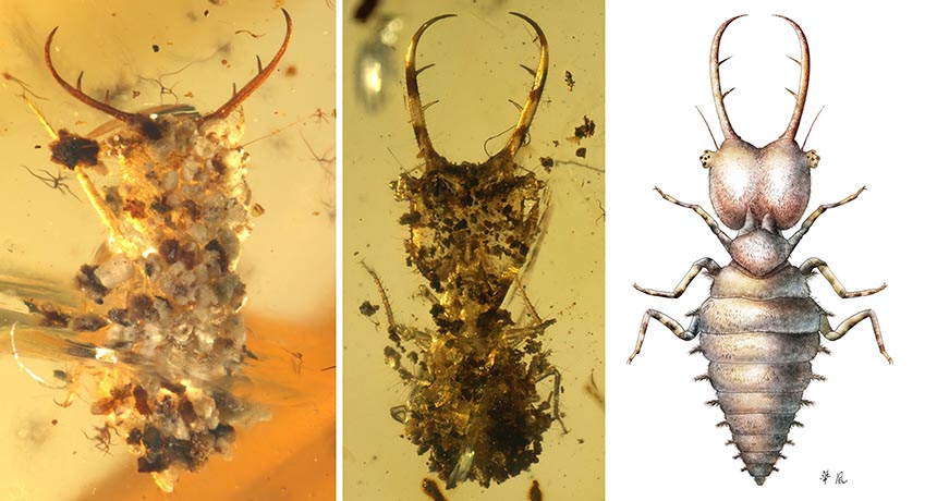
Some insects make dirt look like — well, dirt. And they’ve been doing it for a while.
Donning a bit of debris to blend in with the environment is common practice for a subset of insects and other creepy-crawlies trying to hide from predators. (Crabs, spiders and snails do it, too.) To investigate when this behavior originated, Bo Wang of the Chinese Academy of Sciences and colleagues examined insects preserved in amber from Burma, France and Lebanon that date back 100 million years to the Cretaceous period.
Out of 300,000 insect specimens examined, 39 of them sported what appear to be dirt and vegetation disguises. Anatomical analysis suggests that these insects are early relatives of lacewings, assassin bugs and owlflies. The ancient critters decorated themselves with soil, sand, bits of wood and even tiny ferns, the team reports June 24 in Science Advances.
Until now, only one preserved, dirt-decorated insect from the Mesozoic era had been discovered. But the new finds suggest that this behavior was already widespread in some insect families back then.
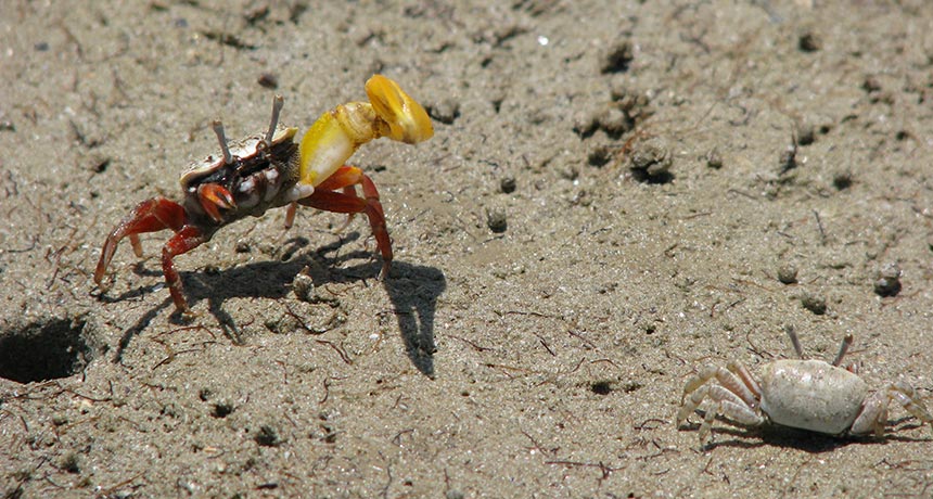
Among people, a man stepping aside to let a woman pass through a door first is seen as a gentlemanly — if a bit old-fashioned — act. Among banana fiddler crabs, though, this behavior is a trap — one that lets a male crab coerce a female into a mating she may not have preferred.
To catch the attention of a female and lure her into his burrow, a male banana fiddler crab stands outside the entrance to his cave and waves the larger of his two claws. A female will look him over and consider his size, the color of his claw and how he’s waving it. If she likes what she sees, she’ll approach him. She might decide to enter his burrow and check it out, and once inside, she might stick around for mating if she thinks that the burrow has the right conditions for rearing her embryos.
When a female approaches a male and his burrow, most males enter first, letting their potential mate follow him down. But many male crabs take another approach, stepping aside and following her into the lair — letting a male trap the female inside and mate with her, researchers report June 15 in PLOS ONE.
Christina Painting of the Australian National University in Canberra and colleagues observed banana fiddler crabs in Darwin, Australia, during two mating seasons, watching what happened as males waved their claws and females made their choice. When a female was interested in a male, the guys entered the burrow first 32 percent of the time. While females were more likely to enter a burrow if a male entered first (71 percent versus only 41 percent when the guy stepped aside), the trapping strategy was more successful in getting a mating out of the meeting. When the male followed the female in, 79 percent of females stuck around the mate. But waiting for her to follow resulted in a pairing only 54 percent of the time.
“The results strongly suggest that entering a male’s burrow first reduces the probability that a female will leave the burrow after sampling it since females are effectively trapped underground in the narrow burrow shaft when the male follows her in,” the researchers write.
So why would a female ever enter a burrow first if there were the possibility that she would be trapped inside and coerced into mating? Perhaps this might give the female a chance to test the male’s strength, the researchers suggest. If she can successfully fight her way out, then the male was obviously not worthy of her attention. Or it is possible that this is just a type of courtship behavior in which no coercion is actually happening. It’s difficult to know exactly what’s going on underground.
This isn’t the first time that the males of a fiddler crab species have been found behaving in what we might consider an ungentlemanly fashion. Males of other species have been found trapping, herding, startling and capturing females in their attempts to coerce a mating. And some male sand bubbler crabs, the researchers note, have even been found behaving somewhat like pirates of the sand-mud flats: Males have been spotted capturing female crabs, carrying them back to their burrows and forcing them into their underground lairs for mating.