Tianjin University sets up first brain-computer interface program to cultivate interdisciplinary personnel
"On August 25, when I logged onto the internet at 10 pm to give a lecture about our new brain-computer interface (BCI) program, 1,200 students and parents were waiting. This clearly demonstrates people's passion for the program, Liu Xiuyun, a professor in biomedical engineering from the Tianjin University (TJU) and director of the TJU School of Pharmaceutical Science and Technology, told the Global Times.
On August 24, the TJU School of Future Technology released an admission notice to the 2024 class of freshmen. Among the majors on offer, there is a biomedical engineering program majoring in the BCI technique, reportedly making the TJU the first Chinese university to off er such a program. The move soon drew the attention of the public on Chinese social media platforms as the country is at a critical stage of the development and application of BCI technologies and devices.
It is not only the first of such college programs in China, but probably the first in the world. It is an experimental project for us to cultivate talent for the future. That is not a small challenge, Xu Minpeng, vice dean of the Academy of Medical Engineering and Translational Medicine of TJU Medical School, told the Global Times.
The applicants from multiple disciplines attended a written examination on August 31, part of them further attended the interview on Sunday, according to Liu. At last, only 20 of them will be selected and officially become the first batch of bachelors majoring in BCI in China.
Technique for tomorrow
As a "revolutionary" technology representing a new form of productive force, the BCI technique aims to establish a direct information pathway between the brain and external devices, achieving so-called control by "thought," thereby revolutionizing human-computer interaction and transforming human production and lifestyle, according to a statement the TJU sent to the Global Times.
BCI is currently a focus of global technological competition and core area for cultivating new tracks, new momentum, and new advantages for future industrial development. In China, the development of BCI technologies and devices has entered a critical period of innovation breakthroughs and application expansion. This necessitates the cultivation of diverse and interdisciplinary talent to meet the demands of the BCI field, providing a continuous driving force for the development of technique, read the statement.
According to the statement, the BCI program is jointly built by the School of Future Technology and Medical School. It brings together advantageous resources from various departments and gathers a top-notch team of BCI talent in China, forming a comprehensive interdisciplinary research and teaching team that covers the entire chain of basic theories, device systems, and transformation in the BCI industry.
The program aims to create an integrated training model through a radiating discipline layout, project-based curriculum system, chain-oriented research training, and a collaborative teaching ecosystem between schools and enterprises. It seeks to cultivate a group of outstanding engineers and scientists who possess the ability to design, manufacture, and develop future bio-intelligent electronic interfaces in the field of bio-information integration, and can lead advancements in brain-computer interaction technology and industrial development, the statement noted.
"BCI is a very complex interdisciplinary field that is connected to, but not limited to, physiology, autopsy, medicine, physics, engineering, computational analysis, automation control, and mechanical design. This is why the new BCI program is put under the School of Future Technology, a platform that is designated to cultivate high-end interdisciplinary talents with strong hands-on skills and innovative capabilities," said Yuan Xubo, the executive vice dean of the School of Future Technology at TJU.
According to Xu, the training plan for the BCI program has significantly established a project-based curriculum that spans all four years of university. In the first year, students will learn the basic concepts of BCI and master simple brain system operations, such as controlling a small ball with their thoughts. In the second and third years, they will progressively grasp principles of neural engineering, analog and digital electronics, automation design, and AI algorithms, while also hands-on creating brain-controlled robots. By the fourth year, students are expected to integrate what they have learned and engage in more systematic paradigm design, algorithm improvement, and application testing aimed at real-world problems.
"This gradual training process aims to develop students' reverse thinking, innovative thinking, practical thinking, and theoretical design thinking step by step," Xu said.
"Through the training, the students will grow into advanced talents with interdisciplinary capabilities. In the future, they will not only be able to enter BCI laboratories to engage in specialized research on BCI, but also participate in the development of high-end medical devices, work in hospitals, or join relevant authorities to take part in the design of policies for related industries," Liu said.
Safe and accurate
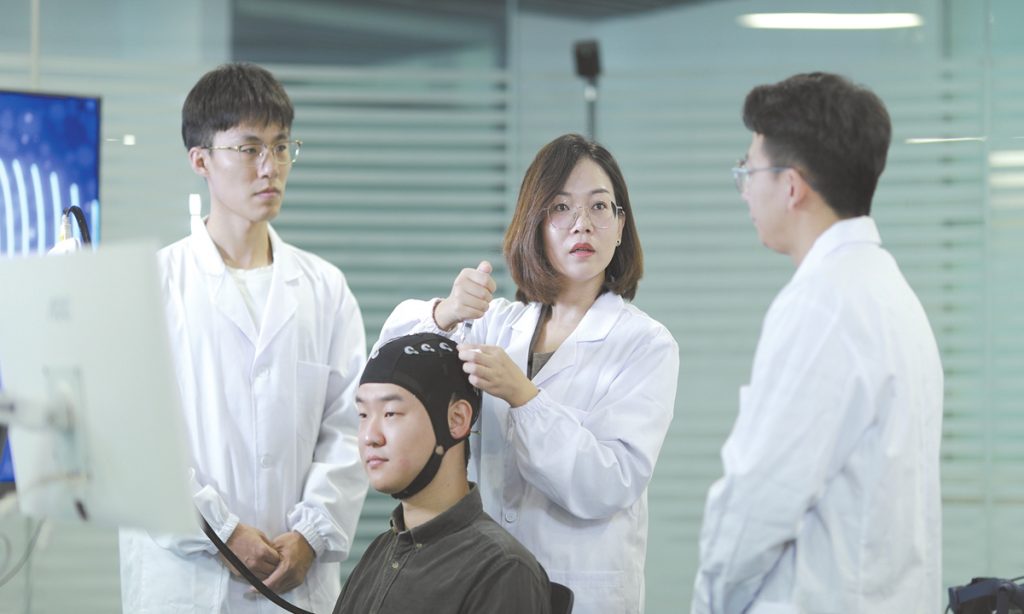
Depending on the electrode position, the methods of BCI can be divided into invasive and non-invasive methods. The invasive method involves implanting electrodes or other devices into the brain or nervous system to achieve a direct connection with the brain. The non-invasive method, on the other hand, uses electrodes placed in the scalp or other areas to record and analyze the brain's electrical signals, thereby establishing an indirect connection with the brain.
Invasive and non-invasive BCI devices have their own advantages and disadvantages as well as applications.
The disadvantages of invasive BCI device include the need for high-risk craniotomy, as well as potential infections, safety concerns, and ethical issues. But it is an effective method for the treatment of severe conditions such as epilepsy. On the other hand, non-invasive BCI device collects comparatively poor-quality signals, but it has a wider range of application, according to Liu Shuang, vice dean of the TJU Medical School and director of the TJU Mental Health Center.
Elon Musk said in January 2024 that his BCI company, Neuralink, had successfully completed the first human brain implantation surgery of its invasive BCI device, called Telepathy, sparking controversy in terms of surgical safety and medical ethics in the world.
Neuralink said in May that a number of wires inside the head of Noland Arbaugh, who is paralyzed from the shoulders down, had been pulled out of position. The company did not specify why the detachment had occurred. Neuralink's implant uses 64 wires to link to the brain; just 15 percent of them were working after the connection severed, The Guardian reported on July 11.
In China the main contributors to the research and development of BCI technologies and devices are universities and hospitals, including the TJU, Tsinghua University, the Shanghai Jiao Tong University, and the Xuanwu Hospital of Capital Medical University in Beijing. Especially in the field of non-invasive BCI, the country is currently taking the lead.
Also in January, a team led by Hong Bo from Tsinghua University announced that they had successfully implanted the world's first wireless minimally invasive BCI device NEO (Neural Electronic Opportunity) into two paralyzed patients' brain for BCI-assisted treatment trials in October and December 2023, respectively.
Hong told the Global Times in a previous interview that compared with Neuralink's technology, the NEO technology has the advantages of higher safety and long-term use.
In the TJU, Liu Xiuyun's team is the first in China to introduce cerebrospinal fluid dynamics assessment technology to help diagnose patients with hydrocephalus. Combined with BCI and proteomics, the team developed a precise diagnostic and intervention technique, which reduces the diagnosis time for patients with hydrocephalus from three days to 30 minutes. So far, the technique has been provided to more than 500 hydrocephalus patients.
Liu Shuang's team has developed the country's first BCI system for screening depression. This system can collect electroencephalogram (EEG) signals from the patient's head to detect brain activity patterns and assess the patient's depressive state, enabling objective screening for depression.
"Normally, the system can generate an auxiliary assessment report on the subject's risk of depression in 10-15 minutes," Liu Shuang said.
The team also developed a technique, the Chirp-based evoked paradigm, that enhances the signal-to-noise ratio of brainwaves gamma oscillations to make the results more accurate and reliable.
Immense potential
On July 6, 1924, German psychiatrist Hans Berger performed the first-ever recording of the EEG on a human, which for the first time allowed the visualization of the electrical activity of the human brain. 100 years later, interaction between human and machine through brainwaves is becoming a reality and such a technique - BCI - is viewed as holding the key to an "era of neuroscience."
The global BCI market size reached $1.5 billion in 2021 and is expected to reach $5.4 billion by 2032, exhibiting a compound annual growth rate of 11.5 percent during 2024-2032, according to market research company IMARC Group.
In early 2016, China launched the 15-year "China Brain Project," or Brain Science and Brain-Like Intelligence Technology, in which the interface played a key role, displaying enormous growth potential. By the end of January 2024, the Chinese Ministry of Industry and Information Technology and six other departments issued the "Implementation Opinions on Promoting Innovation and Development of Future Industries," clearly outlining the goal of creating 10 innovative flagship products, including humanoid robots, quantum computers, BCI, and 6G network equipment.
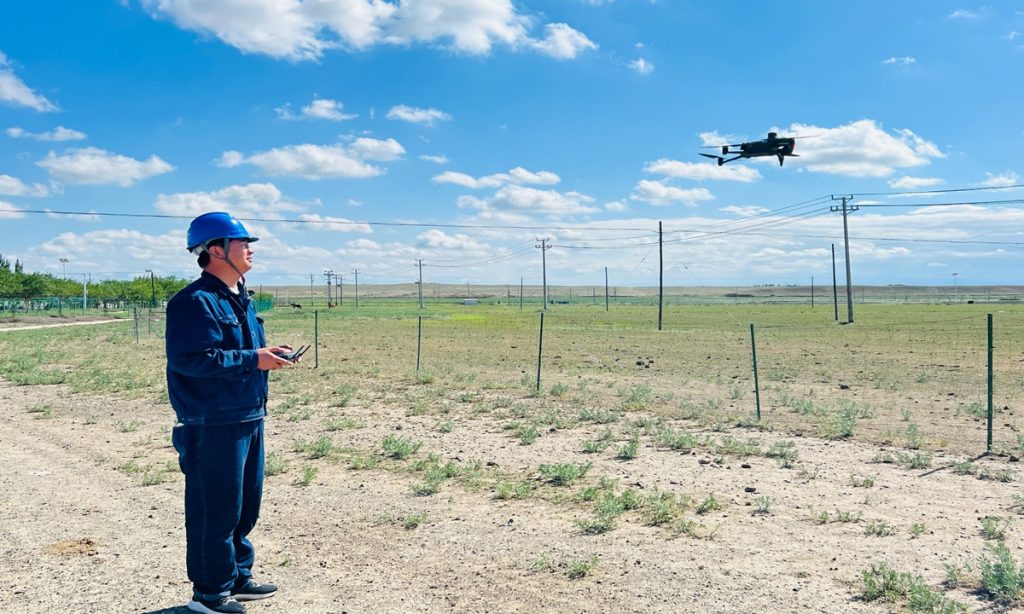
Although mainly used in the medical field, the BCI technique is expected to enter all aspects of people's lives. For example, it can be used to detect whether the driver is fatigued, Liu Xiuyun noted.
TJU scientists have also successfully developed an open-source brain-on-a-chip (BOC) interface system, MetaBOC, which enables a "lab-grown brain" to conduct unmanned control of robots to perform tasks such as obstacle avoidance, tracking, and grasping. MetaBOC represented a significant advancement in the field of BOC technology, offering a versatile platform for exploring the computational mechanisms of biological intelligence, Li Xiaohong, a professor at the Medical School at Tianjin University and the head of the on-chip BCI team, told the Global Times.
BCI is certainly a trend of future technological development. To unlock the immense potential of BCI, it requires a large number of talents to dedicate themselves to the industry. This is why it is necessary to set up a specialized BCI program, according to Xu.
On the other hand, Xu called for patience and caution as it will be a long process to develop safe and high-quality products.